
-

iTero Scanner
Our iTero Scanner is used to provide accurate scans of your teeth for diagnostic models, orthodontic appliances, and virtual treatment planning.
This has all but eliminated the gooey impressions that are used routinely in many other practices. In less than 15 minutes, we can capture the images of your teeth and have you on to whatever is next in your life!
-
Cold Laser Therapy
The cold laser is used after orthodontic appointments to accelerate healing, speed tissue repair, reduce inflammation and discomfort, and to stimulate the growth of new cells
It decreases treatment time and helps start the healing process immediately reducing inflammation and discomfort
*For more information on how Cold Laser Therapy works to help with discomfort and healing, check out these videos on their website: MLS Cold Laser Therapy
-
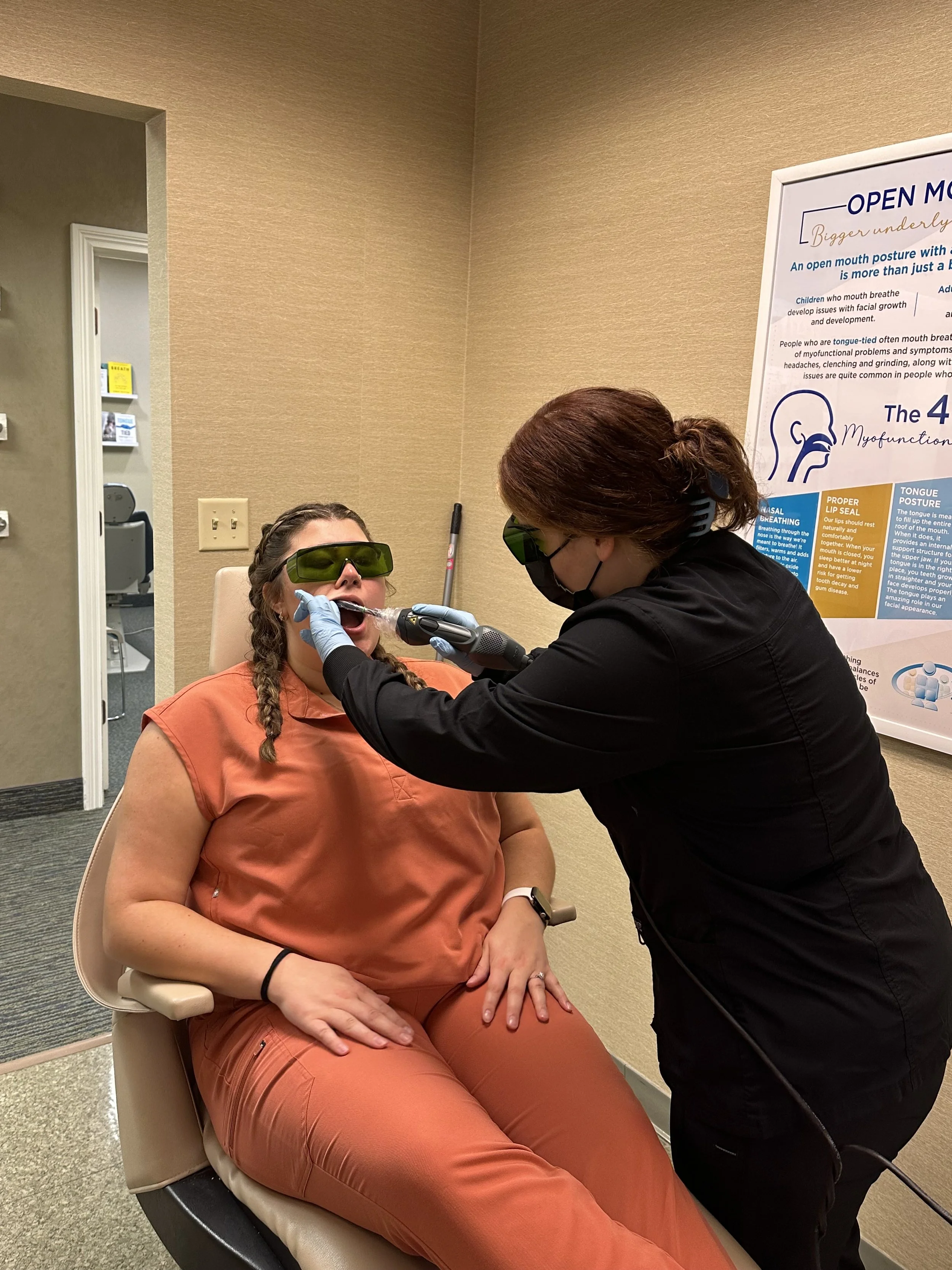
LightScalpel CO2 Laser
Our LightScalpel CO2 Laser is a device that produces a concentrated beam of light. The highly focused CO2 laser beam vaporizes or “erases’ tissue, cleanly and precisely, while sealing blood vessels at the same time.
We use this laser in our office for frenectomies and frenuloplasties (lip and tongue tie releases). The Lightscalpel is ideal for these procedures as it is precise and efficient. Some of the benefits are: shortened procedure time, less swelling and discomfort, reduced risk of infection and scarring, and faster recovery.
*For more information about the LightScalpel laser click here to be taken to the LightScalpel website: LightScalpel CO2 Laser
-

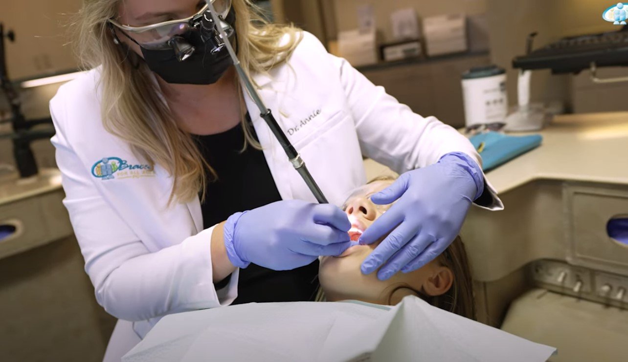
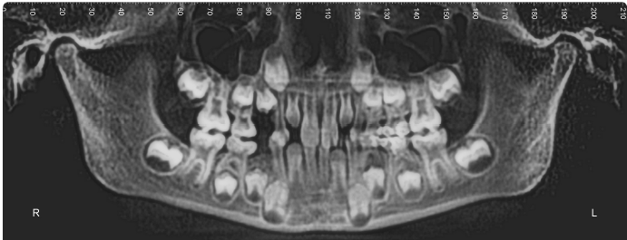
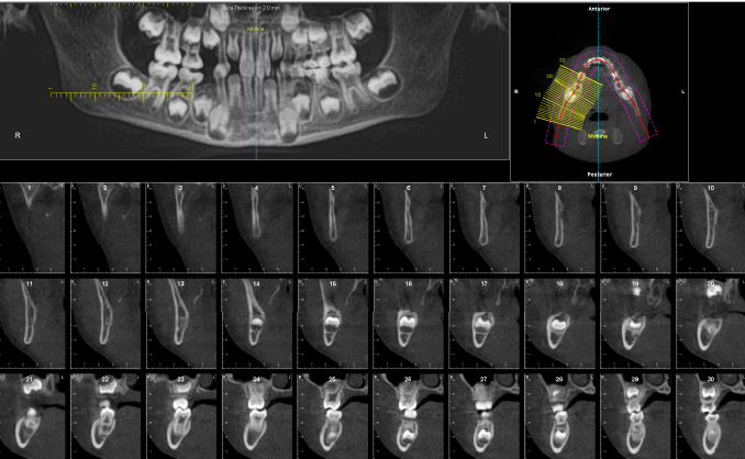
i-CAT 3D Imaging System
Our i-CAT 3D Imaging System provides precisely accurate 3D views to analyze teeth, roots, TMJ, airway, and sinuses without magnification or distortion in less than 5 seconds.
With more information than is rendered from a more traditional x-ray, we are able to provide treatment that is beyond straight teeth.
Use of the i-CAT enables our team of doctors to evaluate the patient’s airway. Constricted airways can cause sleep disorders leading to lack of brain development, learning disorders and attention disorders.
WHAT DO WE SEE WHEN WE LOOK AT YOUR ICAT 3D IMAGE?
DENTAL DEVELOPMENT
In the i-CAT, we are able to see all the primary (baby) and permanent (adult) teeth. We are able to see if there is enough space for the developing teeth to come into proper position. Sometimes, baby teeth that have not fallen out yet may be blocking the adult teeth from erupting. When we see this, we are able to intervene early to make space allowing the teeth to erupt properly.


POSITION OF THE TEETH AND ARCHES
With the images taken from the iCat we are also able to map out the position of the teeth. We are also able to map out the Maxilla(the upper arch) and the Mandible (the lower arch) and compare their relationship to each other. This map can help us determine what appliances will help you achieve your orthodontic treatment goals.

AIRWAY DEVELOPMENT
In the i-CAT image, we are able to see if the airway behind the nose and tongue is developing adequately. If the space is narrowed, a patient may be more prone to sleep disordered breathing and obstructive sleep apnea which can impact growth and development in many ways. When we see a narrow upper airway space we are able to intervene by expanding and protracting the maxilla to make room for the tongue and to open up the airway.
The i-CAT imaging system allows us to take measurements of the Airway Volume. It uses colors to help us visualize airway space, a more restricted airway will have the color red.

*In the i-CAT images below, we are able to see one of our patient cases where they had expansion and protraction with appliances. Here you can see the airway space is much wider in the after picture.


HEAD POSTURE
Ideally, the cervical spine should be curved slightly and the head should be resting above the shoulders. Often, in patients who have narrow airways, the head moves forward to compensate and open the airway. We are able to see the curvature of the upper spine and evaluate head positioning.


TONGUE POSITION
Tongue position helps guide the growth of the upper jaw as well as the development of the nasal passages and upper airway. Ideal tongue posture is the whole tongue resting in the roof of the mouth in a light suction. Although it only captures a moment in time, our i-CAT gives us an idea of where the patient typically rests their tongue.

SINUSES, NASAL TURBINATES, AND NASAL SEPTUM
With our i-CAT Imaging system, we are able to see different sections of the head and neck. Since we are an airway focused Orthodontic practice, we assess the the nasal cavity which contains our sinuses, nasal turbinates, and the nasal septum. Healthy nasal passages are required for nasal breathing which is essential for proper facial and airway development. We can evaluate for potential blockages in the nasal and airway passages and refer to an ENT when needed. Some of the things we look for are:
-Symmetry on both sides of the turbinates and sinuses
-Deviated or “off-centered” nasal septum
-Inflamed/Enlarged or blocked turbinates
-Sinuses that are full or small in size
-Nasal polyps


-TONSILS AND ADENOID SIZE AND LOCATION
A side view of the head allows us to see the size and location of the tonsils and adenoids. When tonsils and/or adenoids are enlarged, the airway is restricted which makes nasal breathing difficult or at times impossible.


TEMPOROMANDIBULAR JOINT (TMJ)
We are able to evaluate the position of the lower jaw in relation to the upper jaw. Often, in patients who have TMJ concerns/pain, the lower jaw is resting further back in the joint space which contributes to the pain and dysfunction. By evaluating the jaw position in the i-CAT we are able to determine if we need to move both the upper and lower jaw forward which helps with joint health.
